Ultrasound during Pregnancy
Ultrasound, also known as sonography, is a method for obtaining images of internal organs by sending high-frequency sound waves into the body. These waves are recorded and displayed as an image in real time.
There is no ionizing radiation in the ultrasound procedure. Obstetric Ultrasound refers to the specialized use of sound waves to visualize and consequently determine the clinical conditions of a pregnant woman and her embryo or fetus.
What are some of the most common uses of this procedure?
Obstetric ultrasound should only be done if it is clinically indicated. Some of the indications include:
- To confirm the presence of a living embryo or fetus.
- To determine the number of embryos/fetuses.
- To determine the pregnancy age.
- To assess the fetus localization (inside or outside the uterus).
- To diagnose fetal abnormalities.
- To assess the placental localization.
- To assess fetal growth and well-being.
- To evaluate the cervix.
How should I prepare for the procedure?
For this examination, you should wear loose clothing, preferably a skirt and blouse. During this procedure, only the lower part of the abdomen needs to be exposed, so if you wear a skirt and blouse you will not be required to remove your clothes.
If the ultrasound is required in the early weeks of pregnancy, you should drink fluids before the procedure because the urinary bladder needs to be full. Air interferes with the sound waves, so if the urinary bladder is stretched, the air area will be displaced and the image of the uterus and embryo will be obtained. In some cases, especially after the first 3 months of pregnancy, a full urinary bladder is not necessary.
An alternative might be the transvaginal ultrasound, especially in the early stages of pregnancy, and for this procedure the urinary bladder should be empty.
What does the equipment look like?
The equipment consists of a probe connected to a monitoring system via a long cord. The probe is a small handheld device that resembles a microphone. The doctor applies a type of lubricating gel to the area to be examined and then gently places the probe on the skin.
The ultrasound image immediately becomes visible on the screen. The obstetrician-gynecologist during the procedure watches the monitor and saves the images. Often, the patient is also able to observe (when the ultrasound office has a second monitor for the patient).
How is the procedure performed?
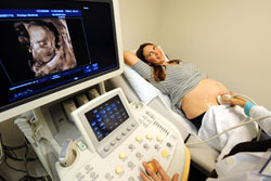
You will be asked to lie on your back and expose the lower part of your abdomen. The doctor will apply a type of gel on your lower abdomen skin.
This gel allows for better transmission of the waves by making it easier to move the probe and by sending the sound bundle directly through the body without interfering with even a small amount of air on the skin. The probe emits high-frequency sound waves while the doctor moves it over your abdomen.
Sometimes the doctor may judge that a transvaginal ultrasound is necessary. This technique is performed like the abdominal ultrasound but the probe is inserted inside the vagina. Through it, often much more detailed and clearer images of the uterus and ovaries are reflected. This technique is very useful in the early stages of pregnancy. For this type of examination, the urinary bladder must be empty. A normal ultrasound examination usually lasts 20 – 60 minutes.
What will I feel during the procedure?
This is a painless procedure. You may feel some slight discomfort due to the pressure the probe exerts on your abdomen, especially when you are asked to keep your urinary bladder full.
Sometimes, the doctor may need to exert pressure with the probe to get closer to the embryo or fetus and to better see its structure. This discomfort is temporary. Also, you may not like the feeling of the gel on your body. With transvaginal ultrasound, this discomfort can be minimal since the probe moves through the vagina.
Who interprets the results and how do I get them?
The obstetrician-gynecologist, who should have the appropriate experience in obstetric ultrasound, analyzes the images and provides the patient with a report of the images' interpretation signed by him.
What are the benefits versus risks?
- Ultrasound does not use X-rays to obtain images – consequently, neither the mother nor the unborn baby are exposed to ionizing radiation.
- Ultrasound has been used to evaluate pregnancy for approximately 4 decades, and there have been no reported cases of harm to patients, embryos, or fetuses. However, ultrasound should only be done when indicated by an obstetrician-gynecologist.

What are the risks?
No adverse side effects have been observed for standard ultrasound examination.
What are the limitations of obstetric ultrasound?
Unfortunately, obstetric ultrasound does not identify all fetal abnormalities. Therefore, if there is suspicion of any possible abnormality, the patient should undergo the invasive test of amniocentesis (evaluation of the fluid in the sac surrounding the embryo) or chorionic villus sampling (evaluation of placental tissue) to determine the health of the fetus.