Another alternative in the treatment of hepatic cystic echinococcosis (Part one)
Until recently, the treatment of liver echinococcosis disease has been considered only surgical. In fact, the spontaneous finding of fully calcified liver cysts indicates that after several stages, the disease has "healed" spontaneously even without any special medical treatment.
Absolutely in this publication, the passive stance in the diagnosis of the disease is not prioritized, nor is the special role of surgery in the treatment of this pathology denied, but by knowing well the development process and the stages of disease evolution, we can successfully use in some cases, less invasive alternatives that are equally successful as the known surgical treatments.
However, of particular interest for very good curative results, remain the attempts to set the diagnosis as soon as possible, in the early stages of the disease evolution.
Ultrasounds are the best, fastest, and least expensive method to diagnose hydatid cysts as well as to follow the progress after treatment.
We are introducing one of the classifications, the one accepted by the World Health Organization, for hepatic hydatid cysts as well as possible treatment alternatives depending on the evolutionary stages of the disease.
Stages according to the WHO classification. Echographic characteristics:
| CL | General cystic lesions, of still undetermined nature. |
| CE1 | Completely watery parasitic cyst, which can be distinguished with difficulty from a simple cyst. (Active). |
| CE2 | Watery parasitic cyst with daughter cysts inside the mother cyst. (Active). |
| CE3a | Parasitic cyst with water and membrane detachment. (Cyst suffering from doubtful activity) |
| CE3b | Solid parasitic cyst but with daughter cysts inside it. (Cyst suffering from doubtful activity) |
| CE4 | Solid parasitic cyst. (Inactive). |
| CE5 | Calcified parasitic cysts. (Inactive). (1). |
 | |
|
Fig. 1. Voluminous hydatid cyst of the first type with visible own membrane in Echo as well as with posteriorly stratified echogenic material. (CE1). Echographic management of hydatid cysts M. Caremani*, L. Lapini*, D. Tacconi*, P. Giorni*, A. Caremani** * U.O. Infectious Diseases - S. Donato Hospital - Arezzo; ** Section of Hematology - S. Donato Hospital – Arezzo. Page 142. Italian Journal of Ultrasound 2005; 8(2): 141-149 | |
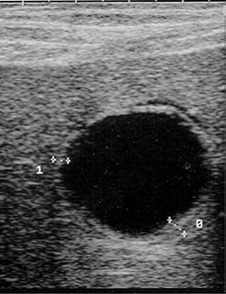 |
Fig. 2. The study of a liver cystic lesion with a 10 MHz probe
Highlighting a proprietary membrane with a thickness of 4 mm with
A triple aspect, typical for the parasitic membrane of a
Hydatid cyst. (CE1). Echographic management of hydatid cysts M. Caremani*, L. Lapini*, D. Tacconi*, P. Giorni*, A. Caremani** * U.O. Infectious Diseases - S. Donato Hospital - Arezzo; ** Section of Hematology - S. Donato Hospital – Arezzo. Page 146. Italian Journal of Ultrasound 2005; 8(2): 141-149 |
The first and second type cysts are evolving cysts with a tendency to grow, while those of the third and fourth type, represent the suffering stage and the involution of the hydatid cyst.
 |
|
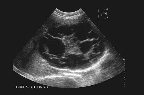
Fig. 3. Hydatid cyst of the right lobe of the liver with a wheel aspect
with daughter cysts inside. (CE2). Echographic management of hydatid cysts M. Caremani*, L. Lapini*, D. Tacconi*, P. Giorni*, A. Caremani** * U.O. Infectious Diseases - S. Donato Hospital - Arezzo; ** Section of Hematology - S. Donato Hospital Page 142. Italian Journal of Ultrasound 2005; 8(2): 141-149 |
 |
|
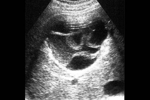
Fig. 4. Hydatid cyst of the liver treated with alcoholization under Ultrasound with the aspect of a swimming flower. (CE3).
Echographic management of hydatid cysts M. Caremani*, L. Lapini*, D. Tacconi*, P. Giorni*, A. Caremani** * U.O. Infectious Diseases - S. Donato Hospital - Arezzo; ** Section of Hematology - S. Donato Hospital S. Donato - Arezzo. Page 143: Italian Journal of Ultrasound 2005; 8(2): 141-149 |
 |
|
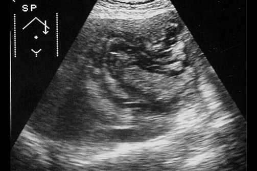
Fig. 5. A large lesion of the third hepatic segment with the aspect of a blade.
Fourth type hydatid cyst. (CE4) .
Echographic management of hydatid cysts M. Caremani*, L. Lapini*, D. Tacconi*, P. Giorni*, A. Caremani** * U.O. Infectious Diseases - S. Donato Hospital - Arezzo; ** Section of Hematology - S. Donato Hospital S. Donato - Arezzo. Page 143: Italian Journal of Ultrasound 2005; 8(2): 141-149 |
 |
|
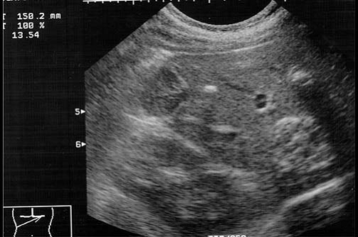
Fig. 6. Subcostal right oblique ultrasound section. Several lesions can be distinguished from which: - One hypoechoic in the 6th segment. - Another hyperechoic in the 7th segment. - And two calcified lesions in the 5th and 6th segments. These are indicators of hydatid cysts in different stages of evolution: (Type CE4 and CE5). Echographic management of hydatid cysts M. Caremani*, L. Lapini*, D. Tacconi*, P. Giorni*, A. Caremani** * U.O. Infectious Diseases - S. Donato Hospital - Arezzo; ** Section of Hematology - S. Donato Hospital S. Donato - Arezzo. Page 143: Italian Journal of Ultrasound 2005; 8(2): 141-149 |
Greetings! I would like to have contact with Dr. Viktor Qershniku because my mother has such a disease. She has been operated on twice within a four-year period and it has recurred again. I would like a consultation from your side. Thank you!
Sent by Monika, më 24 October 2017 në 06:04
Dear Monika,
Can you contact me by phone at
Number 0672161158.
With respect,
Viktori
Replay from Dr. Viktor QERESHNIKU, më 24 October 2017 në 13:13
Hello doctor. Many doctors suspect that my mom has this kind of disease, but the examinations conducted at the American Hospital in Tirana show that she has a cyst with fluid in the liver. She has been treated with medications recommended by the American Hospital and herbal remedies for almost 6 months and there has been no result. Please respond to this case because I am very worried about it. Once again, I am pleading
Sent by Eva Mucmata, më 18 February 2019 në 00:38
Honored ....... ,
I recommend that in your possibility of consulting the patient in Tirana, to send me the Echos made in Kukës and in Tirana as well as all the analyses available, as well as to inform me about the concerns of the patient.
Do you have a dog at home?
With respect,
Viktor QERESHNIKU
Replay from Dr. Viktor QERESHNIKU, më 18 February 2019 në 01:59
Dear Eva,
I recommend that if you have the possibility to consult the patient in Tirana, to send me the Echos done in Kukës and in Tirana as well as all the available analyses, and to inform me about the patient's concerns.
Do you keep a dog at home?
With respect,
Viktor QERESHNIKU
Replay from Dr. Viktor QERESHNIKU, më 18 February 2019 në 02:01
Hello doctor, my father has cysts in his liver, one is 9cm and another is 6cm. They have told him to undergo surgery but my father does not want to do it now but rather in October. I would like to know if there is a possibility that the cysts could rupture and what kind of risk comes from the rupture of a cyst?
Please return an answer.
Thank you!
Sent by Majlinda, më 06 May 2019 në 11:37
Honorable Majlinda,
as you have been informed, I believe, you must be aware that echinococcus cysts are in various stages of development, depending on which the treatment also depends. With as much as you have informed, I cannot give a precise answer on how to proceed.
However, it is important to start treatment with ALBENDAZOLE 2 x 400 Mg. Once daily for 28 days, 14 days break, and then repeat another two cycles.
After these cycles, do a good ultrasound and the antigen analysis.
We can communicate further in the future.
Regards
Replay from Dr. Viktor QERESHNIKU, më 08 May 2019 në 09:38
Hello Dr. Viktori. I am Silvana and my mom has a cyst in her liver. The doctor recommended not to treat it for 6 months. Is it normal not to take any medicine during this time? And can it heal without treatment? With respect, Silvana from Lushnja
Sent by Mikela Xhyra, më 07 July 2019 në 13:23
Dear Silvana,
If it has been proven to be a
CYST OF THE CYSTIC ECHINOCOCCUS OF THE LIVER,
of course, it must be treated first
and followed dynamically.
Best regards.
Respect
Replay from Dr. Viktor QERESHNIKU, më 14 July 2019 në 02:29
Honorable Silvana,
If it has been proven to be a
CYST OF THE LIVER HYDATID CYST,
it certainly needs to be treated first
and followed dynamically.
Past events.
Respects
Replay from Dr. Viktor QERESHNIKU, më 14 July 2019 në 02:30
Hello Doctor,
I have a brother with Echinococcosis and it has progressed, recently it has enlarged to 14 by 12 and I wanted to ask you if there is any treatment without surgical intervention. I have also done a scan and it says that it is a Cystic Echinococcus of the Liver.
How can I proceed in this case because I am very confused and honestly, I don't have much faith in surgical interventions
Sent by Maliq Mici, më 04 Agust 2019 në 23:57
Dear Maliq,
When the diagnosis is confirmed,
Treatment must begin with
ALBENDASOL 2X400 Mg daily for
at least 1 to 2 cycles of 21 days and
Then there are 2 alternatives:
1. Open surgery with a scalpel
2. PAIR technique without opening, which
maybe should be inquired at the American Hospital if they perform it.
It should not be left untreated.
Best regards
Replay from Dr. Viktor QERESHNIKU, më 09 Agust 2019 në 03:59
Dear Maliq,
When the diagnosis is confirmed,
Treatment must begin with
ALBENDAZOLE 2X400 Mg per day for
at least 1 to 2 cycles of 21 days and
Then there are 2 alternatives:
1.: Open surgery with scalpel
2.: PAIR technique without incision, which
maybe should be inquired at the American Hospital if they do it.
It should not be left untreated.
Best regards
Replay from Dr. Viktor QERESHNIKU, më 09 Agust 2019 në 03:59
Where can we find you for a visit, please the address, phone, and do we need to make an appointment?
Sent by Nora, më 14 September 2019 në 19:34
With pleasure, we can communicate
to help you as much
as we can.
I am in Tirana and my phone number
is 0682151598.
Regards,
Viktori
Replay from Dr. Viktor QERESHNIKU, më 15 September 2019 në 10:25