Endoscopic Ultrasonography - EUS (Part One)
What is endoscopic ultrasonography (EUS)?
Endoscopic ultrasonography is a combination of ultrasound and endoscopy to obtain images and information about the digestive tract, its organs, and the surrounding tissues.
Endoscopy is a procedure where a flexible tube with a mini-camera is inserted through the mouth or rectum of the patient to view the digestive tract from the inside. (see: gastroscopy/colonoscopy) while ultrasound uses high-frequency waves to produce images of organs and structures inside the body such as the ovaries, uterus, liver, gallbladder, pancreas, aorta, etc.
Traditionally, ultrasound is done through the skin, after it has been coated with gel, using a probe that acts as a wave transmitter to capture images of organs, but not always have these images been clear.
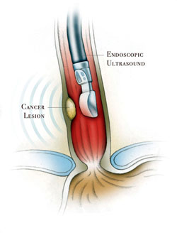
In endoscopic ultrasonography (EUS), a wave transmitter is placed at the tip of the flexible endoscope. By inserting the scope through the mouth into the esophagus and then into the stomach (upper digestive tract endoscopy), high-quality images of the internal organs can be obtained.
Since the wave transmitter is at the tip of the scope, it enables the capture of images in close proximity to the organs, therefore the obtained images are clearer and more detailed than those captured in traditional ultrasound.
Endoscopic ultrasonography can also gather information about the layers of the intestinal wall and the adjacent areas such as lymph nodes and blood vessels. Another use of endoscopic ultrasound is the study of blood vessels using Doppler ultrasonography, and to obtain tissue samples by passing a special needle under ultrasound guidance into enlarged lymph nodes or suspicious tumors. These tissues or cells obtained through the needle are then studied by a histopathologist under a microscope.
When is endoscopic ultrasonography useful?
As it is a new diagnostic method, its applications are still developing, currently it is used for;
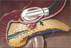
- Staging of cancer of the esophagus, stomach, pancreas, and rectum.
- Staging of lung cancer.
- Evaluation of chronic pancreatitis and pancreatic masses or cysts.
- Study of anomalies of the biliary duct or gallbladder or liver tumors.
- Study of the lower muscles of the rectum and anal canal in evaluating fecal incontinence.
- Study of "submucosal lesions" such as nodules or "bumps" that may hide in the wall of the intestines and are covered by normal mucosa of the digestive tract and which give the impression of something abnormal.
Staging of cancer has become one of the routine reasons for the use of endoscopic ultrasound. The course of patients with cancer depends on the time they are diagnosed, meaning: diagnosing colon cancer in the early stages means that the cancer is only in the layers of the intestine and has not spread to other organs, which gives the patient more chances for a good course of the disease by benefiting greatly.
The opposite occurs when cancer is discovered in advanced stages where it has spread outside the walls of the intestine and to the organs, surrounding lymph nodes, or has spread to distant organs such as the liver and lungs.
Endoscopic ultrasonography gives us complete information regarding the depth and spread of cancer to the organs and surrounding lymph nodes.