Diagnosis (Part Two)
How can doctors identify ACL injuries?
History and physical examination are perhaps the most important ways to diagnose an injured or deficient ACL. In acute injury (sudden), swelling is a good indicator. One way orthopedic surgeons use is that any swelling that occurs within two hours of knee injury usually represents blood in the joint, or a hemarthrosis. If swelling occurs the next day, the fluid is more likely due to an inflammatory response.
Placing a needle in the swollen joint and aspirating (or draining as much fluid as possible) provides relief from swelling and also valuable information for the doctor. If blood is found in the joint, there is a 70% chance that this represents a torn ACL. This fluid can also indicate if the cartilage on the surface of the knee is damaged.
During the physical examination, the doctor will determine how much the ACL is damaged and if other ligaments in the knee or cartilage are also injured.
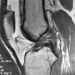
The doctor may order a knee X-ray to rule out the possibility of a fracture. Ligaments and tendons do not appear on X-rays, but bleeding in the joint can result from a fracture, or when parts of its surface are damaged. Magnetic Resonance Imaging (MRI) is perhaps the most accurate test for diagnosing a torn ACL without the need for knee surgery. The MRI device uses magnetic waves instead of X-rays to display the soft tissues of the body. This device creates images that look like slices of the knee. The images show the anatomy and any damage very clearly. This test does not require special dyes or painful procedures.
In some cases, arthroscopy may be used to make a definitive diagnosis if there are suspicions about what is causing a problem in your knee.
Arthroscopy is a procedure that involves inserting a small TV camera with fiber optics into the knee joint, allowing the orthopedic surgeon to see directly inside its structures. The vast majority of torn ACLs are diagnosed without using this type of surgery, although arthroscopy is sometimes used to repair a torn ACL.
Treatment
How do doctors treat ACL injuries?
Non-Surgical Treatment
Initial treatment for an ACL injury focuses on reducing pain and swelling in the knee. Rest and medications such as acetaminophen (Tylenol) can help alleviate these symptoms. You may need to use crutches until you can walk without a limp. Most patients are advised to put normal weight on the knee during walking. The knee may need to be drained with a needle (mentioned earlier) to remove any blood that may be present.
Most patients undergo physical therapy after an ACL injury. Therapists treat swelling and pain using ice, electrical stimulation, and periods of elevation with your foot supported at a height.
Exercises are used to help you regain normal knee and muscle movement. Exercises involving a series of movements should be started immediately with the goal of quickly regaining full knee movement. This includes using a stationary bike, gentle stretching, and careful pressure application on the knee only by the therapist. Exercises are given to improve the strength of the quadriceps muscles and the muscles of the hamstring group (M.biceps femoris, M.semitendinosus, and semimembranosus). When symptoms ease and strength improves, you will be directed to specialized exercises to improve knee stability.
An ACL brace may be suggested. This type of brace is usually custom-made and is not something that can be bought over the counter. It is designed to improve knee stability when the ACL is not functioning properly. An ACL brace is often recommended when the knee is unstable and surgery is not planned. As mentioned, an untreated torn ACL often leads to early knee arthritis. In these cases, doctors prescribe a brace to help prevent knee damage due to a damaged ligament. ACL bracing helps the knee stay in place during moderate activity. However, it may provide a false sense of security and may not always protect the knee during activities that involve jumping, for example. Many orthopedic surgeons recommend wearing a brace for at least a year after surgical reconstruction, so even if you decide to have ACL surgery, a brace is likely a good investment.
Surgical Intervention
If symptoms of instability are not controlled by a brace and rehabilitation program, then surgical intervention may be suggested. The primary goal of the operation is to prevent the tibia from moving too far forward under the femur bone and restore normal knee function.
Even when surgical intervention is necessary, surgeons recommend their patients follow physical therapy for several sessions before the operation. This is done to reduce swelling and ensure that you can lift your knee completely. This practice also reduces the chances of knee pain and can speed up healing after surgery.
Arthroscopic Method
Many surgeons now prefer to reconstruct the torn ACL using a piece of tendon or ligament to replace the torn ACL. This surgery is more often performed with the help of arthroscopy. Incisions are usually made around the knee, but the surgery does not require the surgeon to open the knee. Arthroscopy is used to see inside the knee joint while the surgeon is working. Most ACL surgeries do not require a hospital stay, and many patients return home on the same day as the surgery. Some patients may stay one or two nights in the hospital if necessary.
Patellar Tendon Transplant
One type of transplant used to replace the torn ACL is the patellar tendon. This tendon connects the patella (knee cap) to the tibia. The surgeon removes a strip from the center of the ligament to use as a replacement for the torn ACL.
Transplant of the Muscles of the Posterior Group (M.biceps femoris, M.semitendinosus, and Semimembranosus)
Surgeons may also use the muscles of the posterior group (M.biceps femoris, M.semitendinosus, and Semimembranosus) for the reconstruction of a torn ACL. The transplant material is taken from one of the tendons of these muscles that attaches to the tibia, just below the knee joint. The muscles of the posterior group (M.biceps femoris, M.semitendinosus, and Semimembranosus) extend to the back of the thigh. Their tendons pass through the knee joint and attach on both sides of the tibia. The transplant tendon used in ACL reconstruction is taken from the tendon of the semitendinosus muscle. This tendon runs along the inner part of the thigh and knee. Surgeons also include a tendon attached to the semitendinosus called gracilis as part of the transplant material. When organized into three or four strands, the transplanted tendon has almost the same strength as the patellar tendon used for transplantation.
Allograft Reconstruction (Reconstruction with Foreign Transplant)
Other materials may be used to replace the torn ACL. In some cases, an allograft may be used. An allograft is tissue that comes from someone else. This tissue is taken from the tissues and organs of deceased donors and sent to a tissue bank at the time of death. The tissue is checked for any infection, sterilized, and stored in a refrigerator. When needed, the tissue is ordered by the surgeon and used to replace the torn ACL. The allograft can be from the patellar tendon, the tendons of the muscles of the posterior group (M.biceps femoris, M.semitendinosus, and Semimembranosus), or the Achilles tendon (the tendon that connects the calf muscles to the heel).
Surgeons use the Patellar Tendon as an allograft of the tissues because the tendon comes with the original bone attached to each end of the transplant (from the cup and from the tibia). This makes it easier to adjust the allograft in place. The advantage of using an allograft is that the surgeon does not have to disturb or remove any of the normal tissues from your knee to use as a transplant. The operation also usually takes less time because the transplant does not need to be taken from your knee.
Revision
What to expect after treatment?
Non-Surgical Rehabilitation
Non-surgical rehabilitation for a torn ACL usually lasts six to eight weeks. Therapists apply treatments such as electrical stimulation and ice to reduce pain and swelling. Exercises to improve knee range of motion and strength are gradually added. If the doctor prescribes a brace, the therapist will work with you to get and use it.
You can return to sports activities when your quadriceps and muscles of the posterior group (M.biceps femoris, M.semitendinosus, and Semimembranosus) have regained their full strength and control, you do not have swelling that comes and goes, and you do not have a problem with knee stability.
After Surgical Intervention
Most doctors refer their patients to formal physical therapy after ACL reconstruction. Most likely, you will be involved in a progressive rehabilitation program for 4-6 months after surgery to ensure the best outcome from ACL reconstruction. Initially, the physical therapist meets 2-3 times a week. If the surgery and rehabilitation go as planned in the first six weeks, you may need to follow a home program and meet with your therapist every few weeks for a 4-6 month period.
I received information, but in one line it mentions about the decrease in estrogen, what happens with the weakening of the ligament when we have a decrease due to menopause at the age of 40-50 years. Thank you
Sent by luljetalaci, më 19 Agust 2013 në 11:59
I have an ACL (anterior cruciate ligament) injury. If I undergo surgery, can I play football again?
Sent by erion cara, më 22 December 2013 në 13:10
I have damaged the ACL ligament twice, what needs to be done and if I do not undergo surgery, can I play soccer?
Sent by elsamir, më 28 September 2014 në 04:54