Ear Diseases (Part One)
Non-specific inflammatory diseases of the middle ear and mastoid.
Inflammatory diseases of the middle ear are important first because they are very common and secondly because of the risk of intracranial complications they present as a result of the anatomical proximity between the cavity of the middle ear and the cranial cavity.
Ventilation and drainage disorders of the middle ear.
Pathophysiology. Most diseases of the middle ear are caused by the disruption of two main functions - damage to the function of ventilation of the middle ear and inflammation. Ventilation impairment in relation to the Eustachian tube is based on inflammation of the mucosa in the nasopharynx leading to inflammation of the mucosa of the middle ear.
Pathogenesis. The Eustachian tube does not open regularly during swallowing due to these factors.
- Dysfunction of the levator veli palatine muscle.
- Edema of the tube mucosa caused by chronic inflammations of neighboring structures such as the sinuses, tonsils, or allergy.
- Obstruction of the tube ostium by adenoid hypertrophy in children and adults.
- Infiltration of the tube by nasopharyngeal tumors.
- The middle ear remains unventilated and the remaining air is reabsorbed causing a reduction in pressure in the middle ear serving as a stimulus of the mucosa. In short or persistent blockages of the tube, the reduction in pressure in the middle ear can cause these changes.
- Edema of the mucosa
- Creation of transudate in the middle ear
- Increased resistance in the mobility of the bone chain and retraction of the tympanic membrane
Prolonged obstruction and reduction in pressure in the middle ear can be followed by the following changes.
- Metaplasia of the middle ear mucosa from flat epithelium to ciliated epithelium with cilia and mucus-producing cells.
- Increased secretory activity of mucus-producing cells and mixing of mucus with the transudate created in the ear forming a seromucinous exudate.
- Creation of a cholesterin-containing exudate called cholesterin granuloma.
- The seromucinous exudate and substantial changes that the mucosa undergoes create the ground for a vicious cycle of changes.
Acute blockage of the Eustachian tube.
Clinical presentation. The feeling of pressure in the ear following a situation of rhinopharyngitis accompanied by mild ear pain, feeling of crackling in the nose during swallowing, hearing loss.
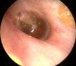
Diagnosis. In otoscopy, retraction of the tympanic membrane, mild hyperemia of the malleus handle, and dilation of blood vessels in the tympanic membrane are observed. If transudate appears in the middle ear, we have a change in the color of the TM and the appearance of the liquid level in pars tensa. Patients complain of mild hearing loss.
Treatment. For treatment, it is necessary to consider the primary cause and its treatment is primary. Rhinopharyngitis is treated with decongestants, antihistamines, vasoconstrictors, to reduce edema in the tube. Antibiotics are started when there are signs of progression of the process towards acute otitis or acute rhinopharyngitis, sinusitis is treated. In case of adenoid hypertrophy, surgical intervention is performed later if necessary.
Chronic seromucinous otitis
Clinical Presentation. The patient feels a sense of pressure and blockage in the ear often following an upper respiratory tract infection and we have hearing loss in one or both ears. We hear noises in the ear during chewing, swallowing, or sneezing. There is no pain.
Pathogenesis. Blockage of the Eustachian tube results in a decrease in intra-tympanic pressure. Obstruction of the tube in the nasopharynx and disorders in the functioning of the tube, especially the inability of the levator veli palatine muscle in cases of cleft palate, viral infections are the most common predisposing causes of the disease.
Diagnosis. In otoscopy, we see a notably retracted tympanic membrane accompanied by small prolapsing areas and the exudate level is observed behind the tympanic membrane. TM is seen discolored and in the external ear canal epidermis produced by the TM is continuously found. In hearing measurement, there is a hearing loss up to 50db for all frequencies giving a flat graphic appearance to the audiogram. The tympanometric curve is flat and compliance is less than 0.2.
In tuning fork tests, we have a negative Rinne. Searching for the primary cause of the infection, we find adenotonsillar hypertrophy, sinusitis, allergy, or tumor.
Treatment. Treatment of the primary disease such as adenotomy, treatment of sinusitis and when the disease continues for more than two months the recommendation is insertion–placement of ventilation tubes in the tympanic membrane.
Acute middle ear otitis.
Begins with the exudative phase lasting 1-2 days and is accompanied by a temperature increase to 39-40 degrees resistant to antipyretics, rarely in children accompanied by meningism. Patients feel strong pulsating pain which is stronger at night than during the day. Patients complain of ear blockage accompanied by noise synchronized with the pulse as well as sensitivity of the mastoid during pressing, in elderly patients the disease often is not accompanied by temperature.
The second phase lasts 3-8 days and is accompanied by rupture of the tympanic membrane and the flow of pus into the external ear canal, at this moment we immediately have the cessation of pain and lowering of temperature. This phase can be shortened by continuous cleaning of the external ear canal accompanied by the use of disinfectants in the external ear canal as well as the use of decongestants from the nose. Early use of antibiotics slightly changes the clinical course of the disease and does not prevent spontaneous perforation of the tympanic membrane.
The third phase lasts 2-4 weeks and is accompanied by the cessation of flows and drying of the external ear canal and the healing of the tympanic membrane.
Pathogenesis.(Routes of infection). The most common route is through the Eustachian tube. The hematogenous route is rare and occurs in cases of infectious diseases such as measles, rubella, scarlet fever, typhus, and septicemia. From the external ear canal, the infection passes into the middle ear by perforating the tympanic membrane or through a previous perforation especially by the entry of contaminated water into the ear. Incorrect methods of removing foreign bodies from the ear can always serve as a cause.
In healthy individuals, the middle ear is sterile if the tympanic membrane is intact.
The infection is monomicrobial and the most common microbes are streptococcus in adult patients, pneumococcus, hemophilus influenza, moraxella catarhalis, and various staphylococci in children. Viral infections prepare the ground for the superimposition of bacterial infection. The infection not only damages the mucosa of the middle ear but also damages the entire internal respiratory system.
Every attack of acute otitis media is accompanied by mastoiditis.
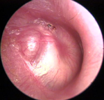
Diagnosis. In the early stages in otoscopy, hyperemia opacity and infiltration of the tympanic membrane are observed, the contours of the malleus handle and the short process disappear. The patient presents conductive hearing loss. In the maximum phase of exudation, the tympanic membrane appears bulging, especially the posterjo-superior quadrant, in this phase the pulsations of the tympanic membrane are seen. The association of otitis with mastoiditis is observed with the appearance of pain when pressing the mastoid retroauricular region.
In the second phase, we have spontaneous rupture of the tympanic membrane and the flow of pus from the tympanic membrane to the external ear canal usually odorless in the form of pulsation. For the verification of the diagnosis and to see the extent of the infection in cases where the infection is accompanied by facial paralysis, loss of consciousness, vertigo, significant neurosensory hearing loss, conventional Ro-graphs, CT helps.
Treatment. Use of antibiotics in high doses 48 hours after the onset of the disease when the situation worsens. Taking cultures for antibiogram in cases where the tympanic membrane is perforated before starting antibiotics. Use of nasopharyngeal decongestants. Use of analgesics. Performing paracentesis in cases where there is pronounced bulging of the tympanic membrane accompanied by high temperature and strong pain.